Lower and Upper Venous Doppler
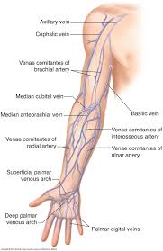
A venous Doppler ultrasound is a diagnostic test used to check the circulation in the large veins in the legs (or sometimes the arms). This exam shows any blockage in the veins by a blood clot or “thrombus” formation.
Upper and Lower Extremity Arterial
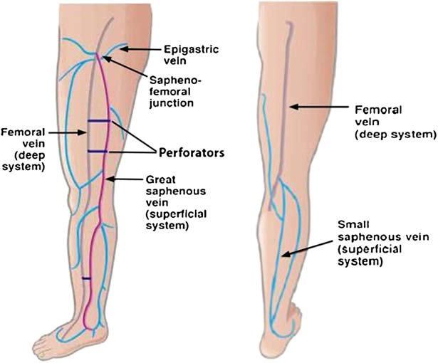
A Venous Doppler test is an ultrasound that uses sound waves to create a picture of your blood flow. The most common use of this test is to search for blood clots. It’s also commonly used for people with varicose veins. Your doctor may order this test if you have leg pain or swelling, or if you have had a previous history of deep vein thrombosis (DVT). Left untreated, these blood clots can break off and pass into your lungs and potentially cause a pulmonary embolism.
Carotid Doppler
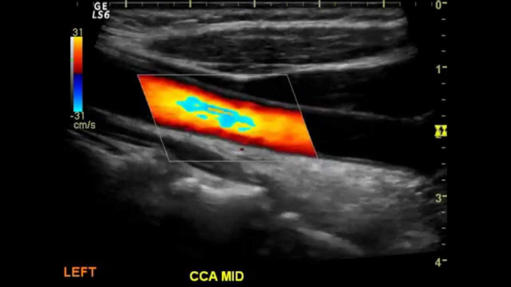
Carotid ultrasound is a safe, painless procedure that uses sound waves to examine the blood flow through the carotid arteries. Your two carotid arteries are located on each side of your neck. They deliver blood from your heart to your brain. Carotid ultrasound tests for blocked or narrowed carotid arteries, which can increase the risk of stroke. The results can help your doctor determine a treatment to lower your stroke risk.
Abdomen Complete/RUQ/Aorta
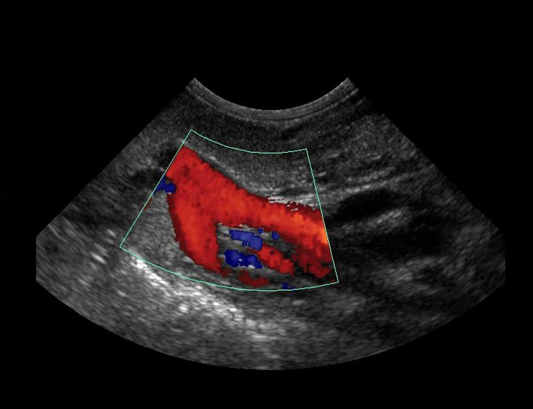
An abdominal ultrasound is done to view structures inside the abdomen. It’s the preferred screening method for an abdominal aortic aneurysm, a weakened, bulging spot in the abdominal aorta — the major blood vessel that supplies blood to the body. However, the imaging test may be used to diagnose or rule out many other health conditions.
Abdomen Doppler
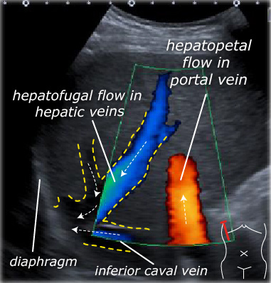
Abdominal Doppler has become an addition in the arsenal of tools available to the physician to aid in the diagnosis of their patients. Abdominal Doppler ultrasound allows ultrasound specialists to evaluate blood flow through the arteries and veins of the abdomen.
Renal and Renal Dopplers
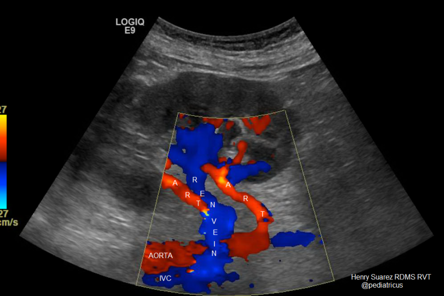
Renal artery ultrasound is a test that shows the renal arteries, the arteries that carry blood to the kidney. These arteries may narrow or become blocked and this may result in kidney failure or high blood pressure (hypertension).
Testicular Ultrasound
A testicular ultrasound (sonogram) is a test that uses reflected sound waves to show a picture of the testicles and scrotum camera.gif. The test can show the long, tightly coiled tube that lies behind each testicle and collects sperm (epididymis).
Thyroid Ultrasound
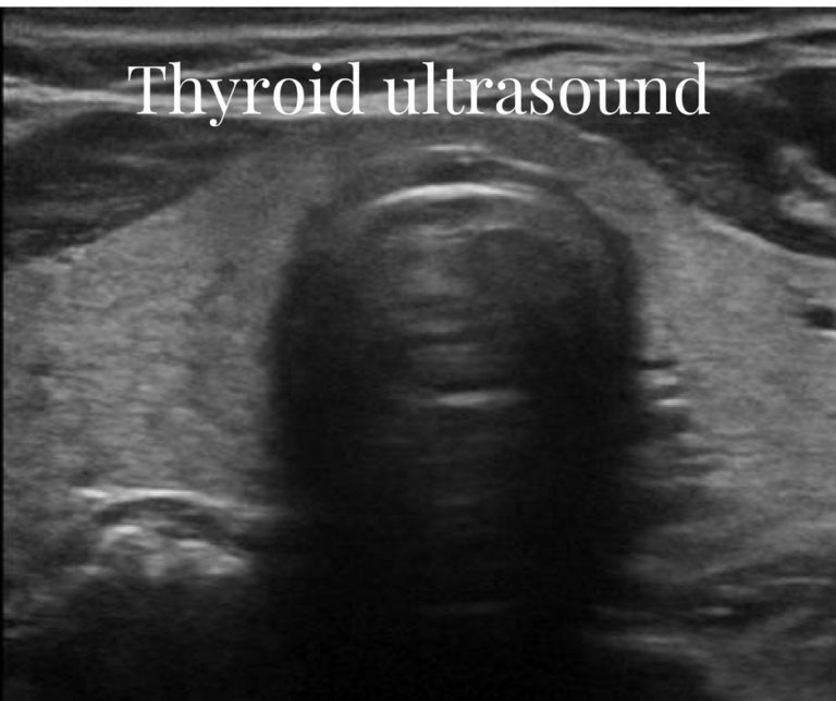
Thyroid ultrasound uses sound waves to produce pictures of the thyroid gland within the neck. It does not use ionizing radiation and is commonly used to evaluate lumps or nodules found during a routine physical or other imaging exam. This procedure requires little to no special preparation. Leave jewelry at home and wear loose, comfortable clothing. You may be asked to wear a gown.
Pelvic Ultrasound
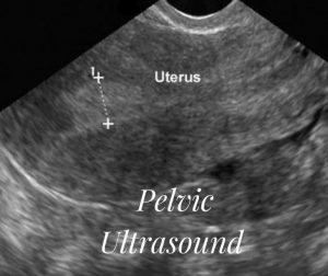
A pelvic ultrasound is a noninvasive diagnostic exam that produces images that are used to assess organs and structures within the female pelvis. A pelvic ultrasound allows quick visualization of the female pelvic organs and structures including the uterus, cervix, vagina, fallopian tubes and ovaries.
Limited OB Ultrasound
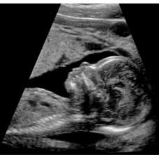
An obstetric ultrasound or sonography is a procedure that uses high-frequency sound waves to produce pictures of a baby inside the mother’s womb. It also shows pictures of the mother’s uterus and ovaries. It is an excellent tool to monitor the well-being of the mother and baby because it is a safe and painless procedure.